A Guide to Glaucoma (Part 3)

Tests and Treatments for Glaucoma
 According to the American Academy of Ophthalmology, glaucoma affects about 1% of people in the United States, and about 10% of people with glaucoma will end up losing their sight.
For some people, no matter how well they live and even if they do everything “right” to prevent it, they will still get glaucoma and develop vision loss. This is why testing your eyes and getting treatment if you have glaucoma is so important.
Tests for Glaucoma
Regular testing for glaucoma is the best way to prevent vision loss and protect your vision. If you wear glasses, glaucoma screening to test if your eye pressure is high may be part of your normal eye exam. Although this screening is good, it is not enough to accurately determine if you have glaucoma.
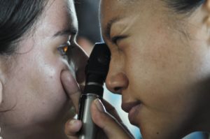 A comprehensive dilated eye exam is the best, most accurate way to tell if you have glaucoma. The American Academy of Ophthalmology recommends that everyone get a comprehensive baseline eye exam at 40 years old. If you are at risk for getting glaucoma (if you are non-white and over 40, have a family history of glaucoma, are diabetic, everyone over 60), then you should get a comprehensive dilated eye exam every 1 to 2 years.
During a comprehensive dilated eye exam, your eye doctor is testing for signs that you have glaucoma, specifically for increased eye pressure, optic nerve damage and loss to your visual field (loss of side or peripheral vision).
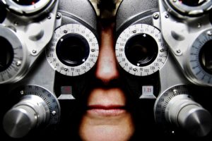 visual acuity test is the most common vision test that uses eye charts to measure how well you see at different distances.”A comprehensive dilated eye exam may include these tests:
Eye Chart (Visual Acuity) Test
A visual acuity test is the most common vision test that uses eye charts to measure how well you see at different distances.
Visual Field (Perimetry) Test
A visual field test measures the vision in your complete field of view to see if you are missing any spots. With a perimetry test you typically look straight ahead at a light and try to see other lights that appear in your peripheral vision (in your side field of view).
A Dilated Eye Exam (Opthalmoscopy)
With a dilated eye exam (ophthalmoscopy) your eye doctor will put drops in your eye to dilate (or open) your pupils. Your eye doctor will then use a special magnifying glass inspect the back of your eye, your retina and optic nerve to check that they are healthy and if there have been any changes to your optic nerve.
During a dilated eye exam, your eye doctor may also use laser scanning, photography or optic nerve and retinal imaging to see the optic nerve inside your eye.
The eye drops used to dilate your eyes may cause your vision to be blurry for a few hours after the exam.
Eye Pressure (Tonometry) Test
In Tonometry, your eye doctor will put drops in your eyes to numb them and then measure the pressure inside your eyes. This test may be measured using the air puff test (non-contact tonometry) or by using an instrument that presses against your eye. Typical eye pressure is around 16 mm Hg (millimeters of mercury).
Cornea Thickness (Pachymetry) Test
In Pachymetry your eye doctor will put drops in your eyes to numb them and then measure the thickness of your cornea using ultrasonic waves. The thickness of your cornea can be related to glaucoma and can affect the pressure in your eye.
Eye Surface (Gonioscopy) Exam
Gonioscopy gives the most accurate diagnosis of the type of glaucoma you may have. In gonioscopy you doctor puts drops in your eyes to numb them and then puts a lens on your eye surface to examine the area that drains eye fluid.
Half of all people living with glaucoma do not know they have this condition. Because few people are aware of glaucoma, most people do not get tested. Getting tested for glaucoma and treatment are the best ways to protect your sight from glaucoma.
Treatments for Glaucoma
 If you have been diagnosed with glaucoma there are many treatments that are proven to be effective in controlling glaucoma and maintaining your vision. Everyone is different and your eye doctor will work with you to find the best treatment for your glaucoma.
Common treatments include medicine, laser and traditional glaucoma surgery. Eye doctors may prescribe any or a combination of these treatments. It is very important that you follow any treatments your doctor prescribes to keep your vision.
Medicine
There are many medicines (both eye drops and pills) that can be used to lower eye pressure. If you have been prescribed medicine for your glaucoma, it is important that you follow the directions and keep taking your medication as directed. You may have to take glaucoma medication for the rest of your life to prevent vision loss.
Eye Drops
Often eye drops are the first step in treating glaucoma. Eye drops can reduce the pressure inside your eyes and can delay or even prevent the start of glaucoma. Researchers from the National Eye Institute found that eye drops to reduce eye pressure helped African Americans reduce their developing glaucoma by 50%.
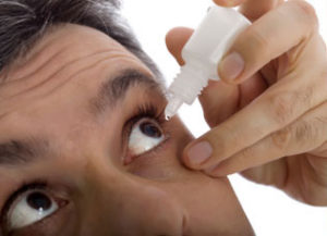 Eye drops work by increasing the flow of fluid from the eye (drainage) and/or by decreasing the production of fluid in the eye. Both of these work to reduce eye pressure.
The best way to take eye drops and to maximize their benefits is to close your eye and press your index finger against the corner of your nose and eyelid (to close your tear duct) for 1 to 2 minutes after dispensing the eye drops. Eye drops can sting or burn for a few seconds when you first use them.
Pills
When eye drops are not enough to control you eye pressure, your eye doctor may prescribe pills to go with your eye drops. Typically these pills reduce the amount of fluid your eyes produce. You may have to take these pills 2 to 4 times a day.
Remember to keep taking your medicine. Quitting or not taking your medicine correctly could lead to permanent eye damage and vision loss. Also, it is important to tell all of your doctors all the medications you take so they can avoid prescribing medicines with dangerous interactions.
Medication and Gene Therapy
Researchers constantly push to find ways to prevent glaucoma and to restore vision loss from glaucoma. A recent study by Boston Children’s Hospital published in Cell journal found a mixture of drugs and gene therapy that could potentially treat glaucoma, restore optic nerve damage and perhaps even to reverse blindness. This study was the first to actually regenerate optic nerve fibers.
Although such studies are promising, they are very new and not yet available to the public. The goal is to find a way to generate new optic nerve fibers using medications and gene therapy to be used in vision recovery.
Laser Glaucoma Surgery
Laser surgery can be effective in treating glaucoma and these treatments may be prescribed for you instead of or along with glaucoma medications. There are many different kinds of laser surgery, and the type you undergo depends on your form and severity of glaucoma.
Laser surgery is typically a day surgery in an out-patient environment (doctor’s office or eye clinic) with the actual procedure typically lasting just 10 to 15 minutes.
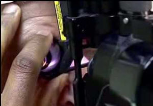 In laser surgery your eye doctor uses a local anesthetic to numb your eye(s). A lens is held to your eye while a laser is used to make small burns (or openings) in your eye tissue. This procedure helps the fluid in your eye to drain more easily, which lowers the pressure. During laser surgery, you may see a bright flash of light.
Your doctor will check your eye pressure 1 to 2 hours after laser surgery. Then typically, you are cleared to go home. You may have irritated eyes and blurry vision after the surgery, but these are temporary. Most people return to their normal daily living activities within a day of glaucoma laser surgery.
Laser surgery is beneficial because there is no cutting of the eye (unlike with traditional glaucoma surgery). However, there are risks, including temporary stinging and inflammation of the eye, temporary increase in eye-pressure (or even eye pressure dropping too low) and a small increase in your risk for developing cataracts.
After laser surgery, you may temporarily need to continue taking medicine to control your eye pressure. However, your eye doctor will monitor this and may reduce your medicines or even stop them altogether in the weeks after your surgery. Other people may still need to keep taking glaucoma medication. It is important to follow your eye doctor’s orders when it comes to taking your glaucoma medication.
Types of Laser Surgery for Glaucoma
Argon Laser Trabeculoplasty (ALT)
Argon Laser Trabeculoplasty (ALT) is used to treat primary open-angle glaucoma (POAG) and is 75% successful in lowering eye pressure. In Argon Laser Trabeculoplasty, a laswer is used on the trabecular meshwork of the eye to open fluid channels and increase drainage. Often an Argon Laser Trabeculoplasty procedure lasts two sessions with half the fluid channels treated each time. This regulates overcorrection and lowers the risk of increased eye pressure immediately after the surgery. Argon Laser Trabeculoplasty can be done 2 to 3 times in each eye.
Selective Laser Trabeculoplasty (SLT)
Selective Laser Trabeculoplasty (SLT) is the most common type of laser surgery and is used to treat primary open-angle glaucoma (POAG) to increase the flow of fluid from the eye, lowering eye pressure. Selective Laser Trabeculoplasty does not cure glaucoma, but can be effective in lowering eye pressure if drops or Argon Laser Trabeculoplasty has not worked.
In this procedure (lasting 1 to 2 sessions), your eye doctor uses a low level laser to make 50 to 100 small burns in the trabecular meshwork (tissue) of your eye. This procedure is called selective because it selectively targets specific cells while leaving other areas of your trabecular meshwork untouched.
Selective Laser Trabeculoplasty is not permanent and more than 50% of people with glaucoma who have had SLT need more treatment within 5 years. You can have Selective Laser Trabeculoplasty twice, but after that it loses its effectiveness.
Micropulse Laser Trabeculoplasty (MLT)
Micropulse Laser Trabeculoplasty (MLT) is a relatively new laser surgery used to treat primary open-angle glaucoma (POAG). Similar to Argon Laser Trabeculoplasty and Selective Laser Trabeculoplasty, Micropulse Laser Trabeculoplasty targets the trabecular meshwork of the eye to increase drainage and lower eye pressure. Micropulse Laser Trabeculoplasty is different in that it delivers short microbursts of energy using a specific diode laser.
Laser Peripheral Iridotomy (LPI)
Laser Peripheral Iridotomy (LPI) is used to treat narrow angle glaucoma or angle closure glaucoma. This type of glaucoma happens when the angle between the cornea and iris are so small that the iris blocks drainage (pupillary blockage) of eye fluid and increases eye pressure. In Laser Peripheral Iridotomy, a lawer is used to make a small hole in the iris to balance pressure in front of and behind theh iris. This causes the iris to back away from the fluid channel so the fluid in the eye can drain (bypassing its normal route).
Laser Transscleral Cyclophotocoagulation
If all other treatments fail to reduce eye pressure, and filtering surgery is not an option, Laser Transscleral Cyclophotocoagulation can be used to treat primary open-angle glaucoma (POAG). In Laser Transscleral Cyclophotocoagulation the eye is numbed using a numbing block to block pain in the eye, while a laser sends energy through the outer sclera into the eye to destroy the some of the ciliary body without harming other eye tissue. The ciliary body is the part of the eye that makes eye fluid (aqueous humor). Using Laser Transscleral Cyclophotocoagulation reduces the fluid produced and lowers eye pressure. This procedure may be repeated more than once.
Laser Endoscopic Cyclophotocoagulation (ECP)
Laser Endoscopic Cyclophotocoagulation (ECP) is similar to Transscleral Cyclophotocoagulation and is used to treat primary open-angle glaucoma (POAG) when eye pressure is high and other options have not worked. In Laser Endoscopic Cyclophotocoagulation, again numbing blocks are used to stop pain while a surgical incision is made so that a laser instrument can be positioned inside the eye and energy is beamed directly to the ciliary body. This also reduces the ciliary body’s ability to make eye fluid and thus lowers eye pressure. As with Transscleral Cyclophotocoagulation, Endoscopic Cyclophotocoagulation can be repeated to control glaucoma.
Traditional Glaucoma Surgery
If you and your eye doctor have tried medications and laser surgery to lower your eye pressure and treat your glaucoma but have been unsuccessful, then you may need traditional glaucoma surgery. There are many different types of glaucoma surgeries that have been proven to be successful in lowering eye pressure.
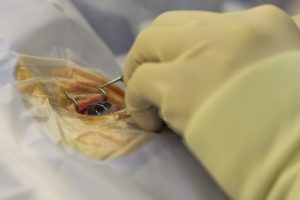 Glaucoma surgery is often a day surgery (that may be in or out-patient) with a number of follow-up visits with your surgeon. Typically, for the 2 to 4 weeks following surgery you should avoid or limit driving, reading, heavy lifting, bending and getting water in your eyes.
You may no longer need glaucoma meds after glaucoma surgery, but be sure to ask your eye doctor and follow their advice before stopping any medications. You also may be temporarily prescribed other medication to reduce inflammation and prevent scarring immediately following surgery.
While traditional glaucoma surgeries have proven to reduce eye pressure and to help guard against optical nerve damage and vision loss, there are some risks. Risks include temporary decrease in vision, bleeding inside the eye, infections and/or leaking at incisions, eye pressure becoming too low and a higher chance of developing cataracts. Also, new drainage ways created by glaucoma surgery can close up, which will again lead to higher eye pressure. Medications prescribed with glaucoma surgery to reduce scarring and inflammation can reduce some of these risks.
Types of Traditional Glaucoma Surgery
Filtering Surgery (Trabeculectomy or Sclerostomy)
Trabeculectomy or filtering surgery is the most common glaucoma surgery and is used to treat both primary open-angle glaucoma (PAOG) and closed-angle glaucoma.
In this type of surgery, a small opening or drainage hole is made in the white part of the eye (sclera) to form a new drainage path for excess fluid to drain out of the eye (reducing eye pressure). As fluid flows through the new hole it creates a bubble or reservoir (called a bleb) where the fluid remains until it is absorbed. The bleb is typically hidden by the upper eyelid so you cannot see it.
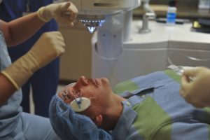 Roughly 50% of people who have filtering surgery no longer need glaucoma medication. Another 35 to 40% still require medication but have better control of their eye pressure. Sometimes, after trabeculectomy or filtering surgery the drainage hole can close which will prevent eye fluid from draining and cause your eye pressure to rise again. Around 15% of people who get a trabeculectomy experience no change in their glaucoma.
Drainage Implant (Valve, Shunt or Seton) Surgery
Drainage Implant Surgery is a good option for reducing eye pressure for people who are not good candidates for or have had a failed filter surgery. With this surgery an implant (valve, shunt or seton) device made from a silicon tube is inserted through the white part of the eye (sclera) into the front part of the eye. The other end of the tube is connected to a plate (or multiple plates) that are stitched onto the surface of the eye and act as a bleb (or reservoir) to hold excess fluid until it is absorbed by surrounding eye tissue. The plate is typically stitched between the eye muscles so that you cannot see it. Drainage Implant Surgery is not as effective in lowering eye pressure as filtering surgery, but it is still a good option.
Canaloplasty
Canaloplasty is a newer, non-penetrating glaucoma surgery that improves the circulation and drainage of eye fluid to reduce eye pressure. Canaloplasty has been called an ocular version of angioplasty because in it your eye surgeon uses a catheter to clear out the drainage canal. Since canaloplasty does not penetrate the eye it is considered safer than filtering surgery, but it is also less effective in reducing eye pressure.
Trabectome, iStent and ExPress
Trabectome and iStent are newer minimally invasive glaucoma surgery procedures that help to lower eye pressure. These procedures are safer than filtering surgeries because they do not use artificial pathways to drain fluid from inside the eye, but rather they bypass any blockage(s) to help eye fluid to drain naturally.
A Trabectome is probe-like device that is inserted into the cornea to send thermal energy into the trabecular meshwork of the eye, increasing the eye’s ability to drain naturally. While this method is safer than filtering surgery, it is also less effective in reducing eye pressure.
The Express is a newer surgical alternative smaller stainless steel shunt device that is placed under the scleral flap in the anterior chamber of the eye. This device lowers eye pressure by redirecting eye fluid from the anterior chamber of the eye. This may reduce risk of infections and surgery complications, but it also is not as effective as filtering surgery in reducing eye pressure.
Although there are many great surgery and medication treatments for glaucoma that reduce eye pressure, even with the best treatments, you can still experience vision loss. It is important to discuss all of your questions and concerns about medication, laser surgery, or traditional eye surgery with your eye doctor.
Some people will continue to gradually lose their vision even though they have done all the “right” things to prevent and treat glaucoma. This is why it is so important to regularly get your eyes tested and to carefully work with your eye doctor to monitor and treat glaucoma.
Early detection and treatment can be your best defense against vision loss. The greatest way to protect your vision from glaucoma is to get your eyes checked. January is a good time each year to do this because it is Glaucoma Awareness Month.
If you do have vision loss from glaucoma, there are many amazing low vision aids and blindness aids to help you live a full, independent life. We will discuss low vision and blindness technology for glaucoma in Part 4 of this series.
As part of our work to increase awareness about glaucoma and glaucoma-related vision loss and living with glaucoma we have written a 4-part Guide to Glaucoma. Please share this Guide with friends and remember to get your eyes tested for glaucoma annually. If you do have glaucoma-related vision loss, call us at 888-211-6933 to learn how low vision aids can help you.
In Part One of our series “A Guide to Glaucoma” we discussed what glaucoma is, the warning signs and symptoms of glaucoma and the stages of glaucoma-related vision loss.
In Part Two of our Glaucoma Guide we discussed who’s at risk for developing glaucoma and how to prevent vision loss from this disease.
In Part Four of our Glaucoma Guide we discuss how glaucoma affects your vision and low vision and blindness aids for people with vision loss from glaucoma.
Please share this important information with your friends and family, encourage everyone you know to get a comprehensive eye exam annually, and be sure to check back for Part Four of our Guide to Glaucoma to learn about the different Low Vision and Blindness Aids for People with Vision Loss From Glaucoma.

